Anchiornis: Putting skin on the bones...with science!
/
For paleoartists (and the scientists who work with them), properly restoring the skeleton is merely the beginning of figuring out how a dinosaur looked in life, since muscles, tendons, and other soft tissues that make up the final shape of an animal can be quite different than the skeleton alone. This requires an extensive knowledge of comparative anatomy from living organisms, a technique which has become more formalized in recent years with the application of Extant Phylogenetic Bracketing (EPB), which leverages the work of phylogenetics (the science of who is related to whom) to test inferences about muscle origins and insertions, and other soft-tissue hypotheses.
When we are really lucky these hypotheses can be tested directly, such as when we find extensive soft-tissue preservations of a fossil like the "mummified" hadrosaurs. For stem birds we have had an embarrassment of riches when it comes to their epidermal coverings (fur, feathers and other fluff), but their fuzziness has generally covered up any information that might have been preserved about the shape of the underlying soft-tissues, or about the non-feathered skin structures.

That's all about to change. In a paper published today by my colleagues and I, dozens of Anchiornis specimens were studied using laser-stimulated fluorescence (LSF), a technique that can reveal preserved soft-tissue details that are not clearly evident in normal lighting conditions. Although no one specimen revealed an entire outline, by cobbling together the best of the dozen or so specimens with LSF-revealed skin tissues we now have a much better idea of what Anchiornis looked like, as well as a good test of how well EPB works. For paleoartists, and those who simply want to know exactly what a dinosaur looked like when alive, this clarifies many important details of the skin and muscular anatomy.
Much will be (rightfully!) written around the web on how laser-fluorescence works, or how these findings impact hypotheses on the origin of birds and flight, but for this article I'm going to concentrate on what the paper tells us about the anatomy and appearance of Anchiornis.
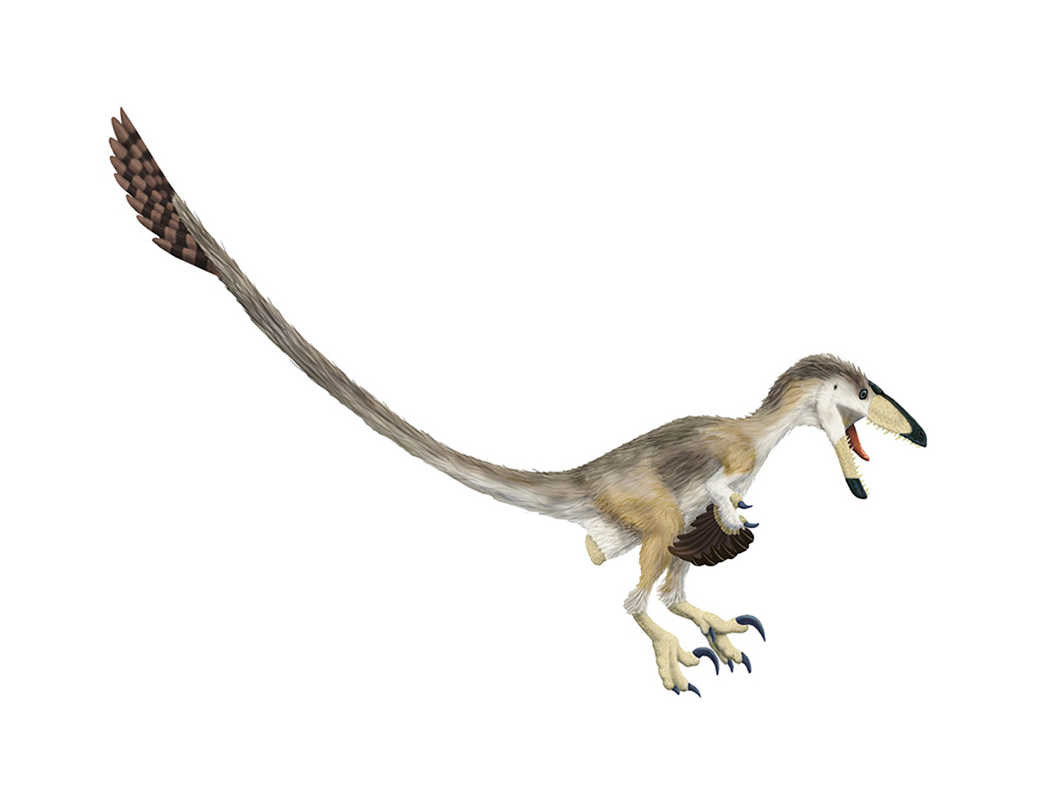
About Anchiornis: If Anchiornis is not a familiar dinosaur to you, you should know that it's a small paravian theropod dinosaur - that is, it's closely related to troodontids, dromaeosaurs, and the lineage that lead to birds. I can't be more precise than this, because exactly which of those three branches it's on has been the subject of some debate and does not yet have a consensus view. But given it's location we would have already known that it was a feathered and winged theropod.
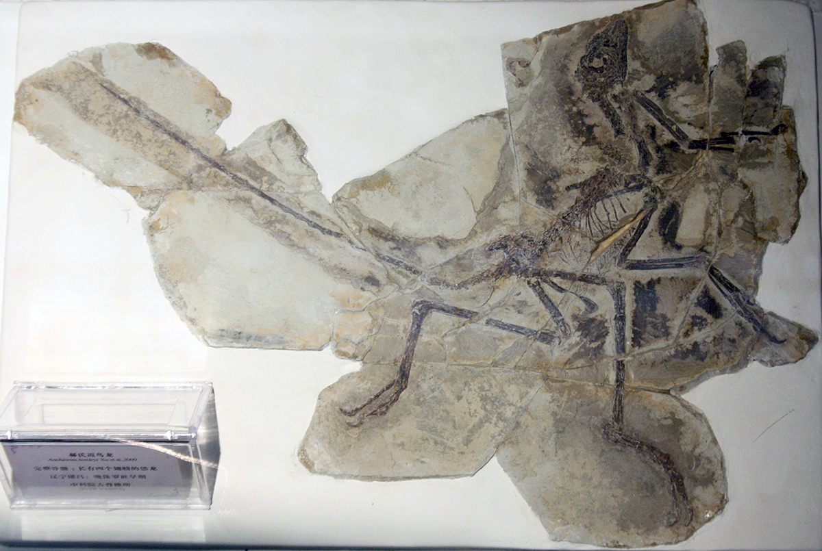
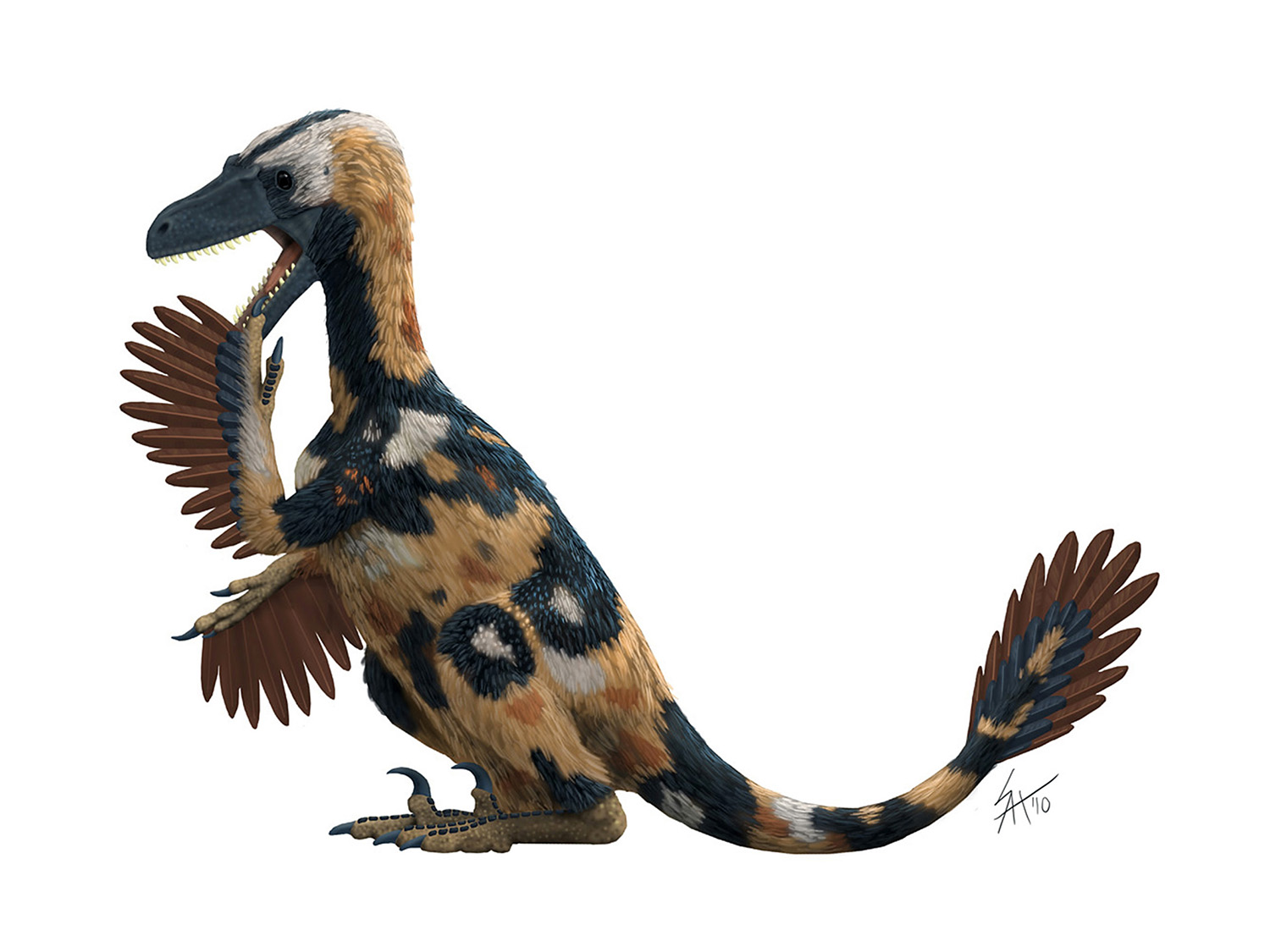
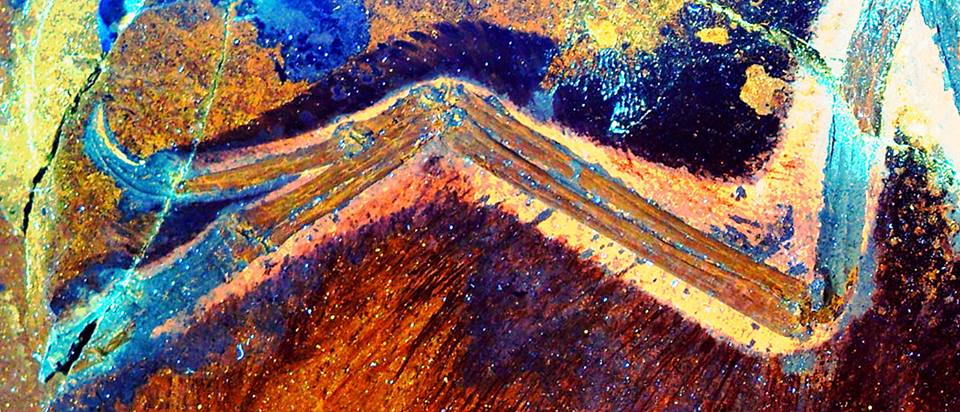
A lovely specimen of Anchiornis, taken by David Hone.
We don't have to assume this, however, as early specimens of Anchiornis were found with feather impressions, including branching feathers on the hind legs. This sometime leads people to refer to Anchiornis as a four-winged theropod, sort of like Microraptor, but it should be noted that the feathers on the hind legs of Anchiornis are not nearly as large or "wing-like" as what we see in Microraptor. The feathers were exquisitely preserved, however, right down to the distribution of melanosomes, the cellular organelles that determine the color of a feather. As a result, Anchiornis has the designation of being the first non-avian dinosaur to have its color scheme scientifically reconstructed.
OK, so what's new already? With so much awesomeness already known for one dinosaur, you might be wondering what could be left to find? As it turns out quite a lot! Using the laser-fluorescence technique developed by Tom Kaye, dozens of specimens were scanned at the Shandong Tianyu Museum of Nature, and many of them revealed details of skin and scales that otherwise would have been missed. Take this image for instance:
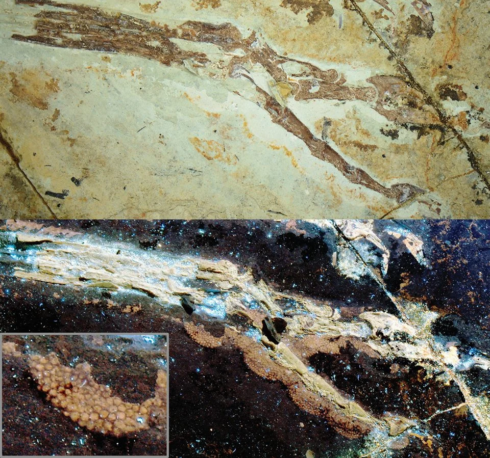
The foot of Anchiornis. Laser-FLUORESCENCE images on the bottom.
First of all, let me take a moment to point out how ludicrously awesome that bottom image is. You can see the scales on all of the foot pads! Notice on top of metatarsals on the upper left you can see what appear to be enlarged scales like you see in many living birds. Unfortunately they are minimally exposed in this specimen, so it's sadly equivocal as to what is going on. There are also some scales found up on the tibia in other specimens, suggesting an interesting interplay of scales and feathers on the hind limbs.
Some other specimens also showed the full extent of the keratinous sheaths on the hand and toe claws. I was particularly pleased to see that the toe pads overlapped the joints between each toe bone - exactly the way we have inferred based on trackway evidence and comparisons with living dinosaurs (you know, birds).
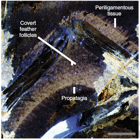
Another image making the media rounds shows off some skin details from the arm:
Here you can see the claw sheath on the thumb claw. You can also see how thick the soft-tissue is on the front (palmar) side of the thumb. In retrospect this makes sense given how large the tendon insertion is on the thumb claw, but it's something that is almost always underemphasized in skeletal and life reconstructions. Another surprise is the extent of the patagium (skin flap) in front of the elbow. This feature is seen in living birds (you buffalo wing fans have eaten your fair share of it), but it had usually been assumed to have evolved in dinosaurs closer to birds - those that were capable fliers. Now we will have to reassess the role of the patagium on the arm (since there is a tendon involved it plays a role in increased leverage) pre-flight. And artists will need to consider expanding the animals they restore with a patagium, potentially including at least all paravians, and (more speculatively) perhaps all winged theropods.
Here is the take-home figure from the paper:

When looking at this figure, the colored portions of the skeletal correlate with the color at the top of the photos - indicating the specimen that best showed off the shape of the skin underneath. The first thing I should note is that strictly-speaking, this figure (and the skeletal at the top of the page) are not showing the same sort of information as the other skeletal drawings on the site. Normally my skeletal reconstructions are done without skin or scale depth added - they are basically flayed. The Anchiornis skeletals here do incorporate skin depth wherever possible.
There are many topics worth covering, but as this is already a giant wall O'text I'll just hit on the major topics that artists and dinosaur-fans should take note of:
Drumsticks: For quite a while paleoartists "in the know" have been illustrating theropod knees with large, avian-style drumsticks. STM-133 and STM-118 shows this inference is correct, as both show calve muscles that rapidly increase in size as you move up the shin towards the knee.
Fat ankles: Several specimens, including STM-133 show a surprising amount of soft-tissue in front of the ankle joint. This certainly wouldn't be due to muscle mass, but instead some combination of tendons and connective tissues seems to pad it out, and suggests that theropod ankles (and maybe all dinosaur ankles?) should be fatter than they are normally illustrated. Likewise, many skeletal reconstructions show the skin hugging the long ankle bones (the metatarsals), but several specimens (including STM-147 and STM-114) show a lot of tissue on the back side of the metatarsals, where thick tendons must have run from the drumstick muscles down to operate the feet.
The pubis: There has often been debate over how to restore the large, sometimes-retroverted pubic boot of paravians. STM-118 preserves a clear outline of the lower body cavity around the pelvic region, and though it suggests a thicker pad of tissue under the boot than most skeletal restorations show, it none the less shows the body largely following the shape of the boot, not covering it up in giant mounds of flesh. Also note that after the boot (on the posterior side) the body margin turns upwards at almost a right angle, continuing up towards the ischium rather than completely burying it.
The tail base: While the skin from the pubis does not extend onto the tail, there is a significant belly of soft-tissue extending from the ischium to the base of the tail. So illustrations that show the ischium sticking out, or the skin being shrink-wrapped right from the ischium to the tail are not correct for Anchiornis, and given the information from hadrosaur "mummies" I'd wager it's not likely for any dinosaurs.
We're actually not too bad at this anatomical reconstructing thing: There have been many cautionary warnings about what we know and don't know about restoring extinct animals. They are still quite valid, and you'll notice that there are several important takeaways from this study that suggest I'll have to spend time revising some of my skeletals (especially the elbows and ankles) that I'd done before I started working on this project. At the same time, when it comes to the nuts and bolts of muscles and tendons I think we should be rather pleased with ourselves - despite the inferential nature of the work we're clearly on the right track. And further discoveries of this kind can only help us further test our phylogenetically bracketed hypotheses.
Check out that non-scaly skin under the feathers!
I know I for one cannot wait!
P.S. If there's interest in a particular topic (covered above or just something you notice in the paper or image) let me know in the comments below and I'll try to do a follow up post.